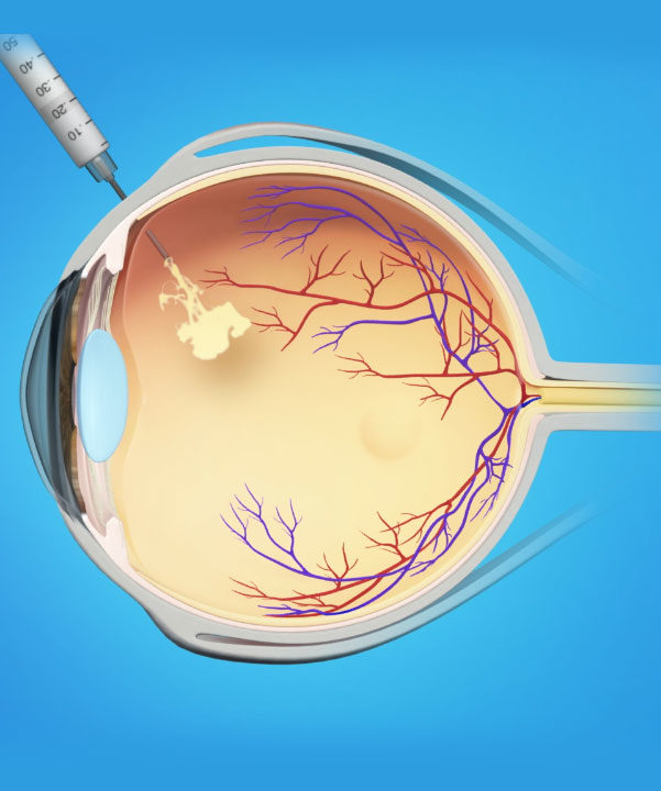
Injections
September 27, 2019
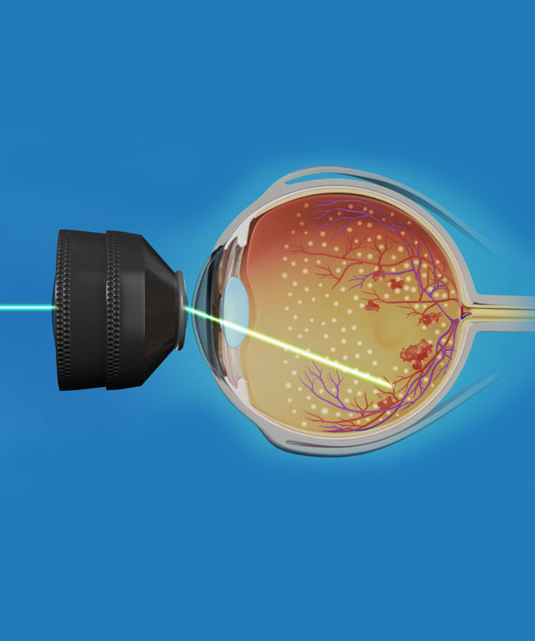
Laser Surgery
September 27, 2019Eye Imaging refers to digital scans or photographs (no radiation involved) of the back of the eye that provide information often not apparent with a detailed examination alone. The images help us improve diagnosis and allow us to best prevent and treat potentially blinding conditions as early as possible.
We at Manhattan Retina and Eye use some of the most cutting edge technology available while still providing individualized care and patient education.
Our imaging machines are capable of taking 100,000 scans per second with HD imaging detail and wider field view. Which helps us to provide the best diagnostics for the patients.
Imaging Machines We Use
Who Gets Eye Imaging
Usually, people with the following diseases or conditions require eye imaging.
Diabetes: Angiography and OCT imaging can detect vision-threatening new blood vessels forming and swelling in the retina. Diabetic Retinopathy is a leading cause of blindness in the US with the vast majority of vision loss being preventable.
Macular degeneration:
The centre of your retina gets affected and your most functional vision can be affected.
“Dry” macular degeneration is the most common form of this disease (More than 95% of the cases).
“Wet” Macular Degeneration: Abnormal blood vessels growing under the retina start to bleed and leak fluid and material that can damage the retina.
Glaucoma: Damage to the optic nerve that is often asymptomatic at first but a leading cause of visual impairment globally.
Retinal Vein Occlusions: Tiny Blood vessels in the eye can be blocked and lead to swelling of the retina or abnormal blood vessel growth.
Retinal Toxicity: The arthritis drug hydroxychloroquine (Plaquenil) can damage your retina.
Many more retinal conditions can affect your retina and threaten your sight. Proper clinical exam and diagnostic imaging are essential to prevent vision loss and improve vision.
Related posts


