About Dr. Arnold D Yagoda
Retina Specialist Board Certified by the American Board of Ophthalmology
Staff at New York Eye and Ear, Mount Sinai, and Lenox Hill
Dr. Yagoda has been in practice as a leading retina specialist since the 1980s.
M.D. from Cornell University
Retina Fellowship: Montefiore/Albert Einstein College of Medicine.
Ophthalmology Training: Lenox Hill Hospital
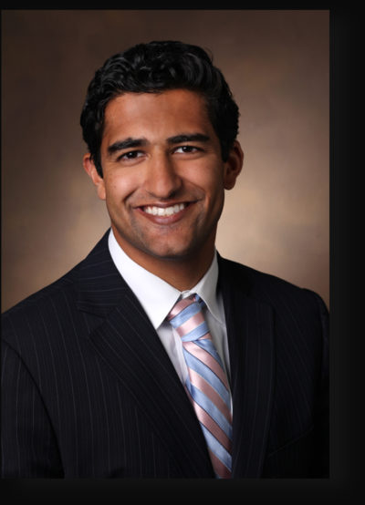
About Dr. Ravi Parikh MD MPH
Retina Specialist Board Certified by the American Board of Ophthalmology
Retina Fellowship Training with Harvard Medical School/Massachusetts General Hospital/Massachusetts Eye and Ear
Vitreous Retina Macula Consultants of New York (Preceptors K Bailey Freund and Lawrence Yannuzzi MD)
General Ophthalmology Training from Yale University/Yale-New Haven Health
M.D. Vanderbilt University as a Canby Robinson Scholar (Full-Tuition Merit Scholarship for Leadership and Academic Excellence)
M.P.H. Harvard University in Health Policy and Management
Groundbreaking Research
Cutting-Edge Diagnostics
World-Renowned
Improve your Vision with
Our Eye Services
ZEISS CIRRUS 6000
Make every second count with high-performance OCT.
Performance OCT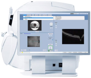
100,000 scans per second to power your practice
CIRRUS® 6000 is the next-generation OCT from ZEISS, delivering high-speed image capture with HD imaging detail and a wider field of view so you can make more informed decisions and spend more time with the patients who need it.
Learn how you can maximize patient throughput and practice efficiency with ZEISS CIRRUS 6000.
Faster, wider with a new level of detail
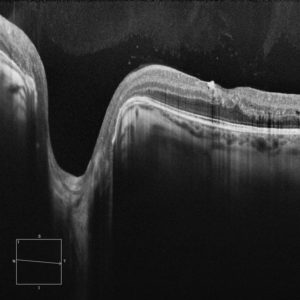 At 100,000 scans per second, ZEISS CIRRUS 6000 enables clinicians to image a larger field of view up to 12mm in a single scan. It also captures high-defintion (HD) OCT and OCT Angiography (OCTA) scans, revealing the finer microvascular details of the retina and providing more insight into your patient’s condition.
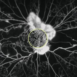 Making the revolutionary, routine.
ZEISS AngioPlex OCT Angiography
AngioPlex® OCT Angiography from ZEISS ushers in a new era of eye care with non-invasive imaging of retinal microvasculature—taking glaucoma and retinal disease management and treatment planning to the next level. By offering the industry’s most comprehensive tools for assessing and analyzing a range of pathologies, ZEISS provides a complete OCT Angiography (OCTA) solution.
Proven analytics
CIRRUS-powered treatment decisions
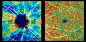 As the pioneering OCT technology, the CIRRUS platform offers clinicians extensive, clinically-validated applications—for retina, glaucoma and anterior segment—that allow for precise analysis, faster throughput, and smarter decision-making across a range of clinical conditions and patient types.
- Macular change analysis lets you track change between visits with confidence
- Glaucoma: Guided progression analysis and comprehensive tools for glaucoma management
- Epithelial thickness mapping, widefield HD corneal imaging, and more
- AngioPlex Metrix: OCTA quantification tools for retina and glaucoma
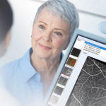
Patient-first design
Unique patient-centric platform designed for the future.
With ZEISS CIRRUS, your patient data is never left behind. CIRRUS is the platform that allows seamless transfer of raw, dynamic patient data from previous generations, allowing clinicians to manage their patients with utmost care, across generations.
Introducing California – Our latest Ultra-Widefield Retinal Imaging Device
Designed specifically for ophthalmologists and vitreo-retinal specialists, our California is the latest addition to our ultra-widefield retinal imaging device technology lineup. With new hardware and software, we are helping you see more, discover more and treat more effectively to promote better patient outcomes.
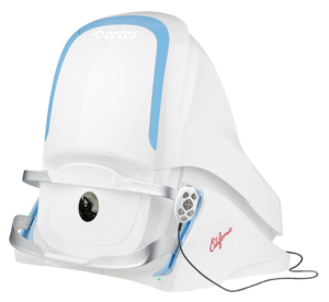
Why California?
We are committed to further strengthening our clinical evidence while demonstrating the importance of imaging the entire retina. California includes a new UWF optomap® imaging modality; Indocyanine Green angiography (icg) while retaining:
- -Composite color
- -Red-free
- -Autofluorescence (af)
- -Fluorescein angiography (fa)
Images are now presented in ProView which displays optomap in a consistent geometry that accurately represents anatomical features in the retina. Further, ProView enables automatic image registration for disease tracking over time, and inter-modality image comparison.
New proprietary optical hardware optimizes and maintains resolution of the optomap images throughout the scan of the retina resulting in more clarity in the far periphery.
Image overlay enables comparison between composite color images and red-free, af, fa, or icg images. Additionally, comparisons can be made between different images or different dates by scrolling through all stored images.
Benefits of California
In addition to the benefits found with all of the UWF devices from Optos such as 200 degrees or up to 82% of the retina captured in a single image, in multiple modalities, as well as eyecare professionals being able to see 50% more of the retina when compared to other conventional imaging devices, California offers the following benefits:
- Compact to reduce space requirements.
- New design leads to ease of use and faster image capture.
- Non-mydriatic high-resolution imaging through many cataracts and/or 2mm pupils saves time in busy practices.
- Browser-based image review enables simple integration and easy access to your data from any connected pc or tablet in a HIPAA compliant environment.
- Interweaved angiography enables parallel capture of fa and icg images without manually switching between imaging modalities.
Optos would like to invite you to learn more about the California and contact us if you have questions or wish to partner with us to help your practice.


